MRI’s, Scans and X-ray
MRI
Magnetic Resonance Imaging (MRI) combines a powerful magnetic field with an advanced computer system and radio waves to produce accurate, detailed pictures of organs, soft tissues, bone and other internal body structures. Differences between normal and abnormal tissue is often clearer on an MRI than CT. There is no radiation exposure with MRI machines.
An MRI scan will take 30 minutes to an hour. With MRI’s images are taken as cross sections of the body. Patients must remain perfectly still through an MRI scan. The machine generally makes loud knocking sounds. A comprehensive patient screening procedure is followed as due to the magnetic field, special precautions are made or exams may be canceled for patients with cardiac pacemakers, tattoos and metal implants. There is a weight limit for the MRI table. There is also an opening a very large person may not be able to fit into.
- Imaging soft tissue, organs an internal structures
- Will show abnormal and normal tissue
- no radiation used

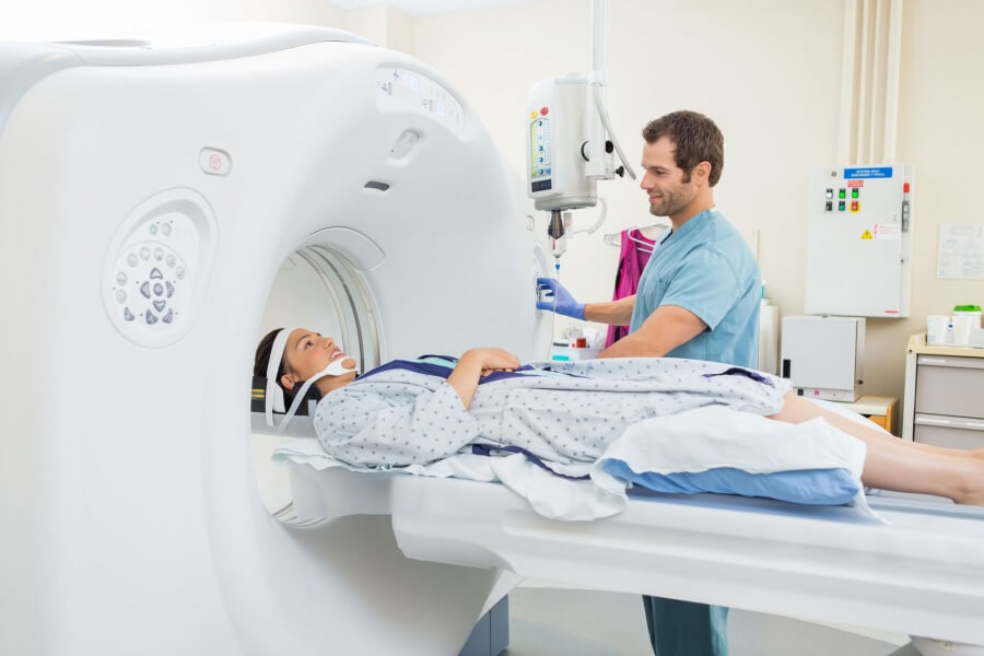
CT / CAT Scan
Computed (Axial) Tomography (CT or CAT scan) commonly a 5-20 minute painless exam that combines the power of X-rays with computers to produce 360 degree, cross-sectional views of your body. CT is able to image soft tissue, bones and blood vessels all at once. It provides the radiologist with details of bony structures or injuries.
The machine has a tube design but is slim from front to back and is comfortable. A large person may not fit into the opening of the CT scanner or may be over the weight limit for the moving table. While it is a painless process, there is radiation exposure with CT scans.
- Imaging soft tissue, bone and blood vessels at once
- Pinpointing issues with bony structures (which are commonly injuries)
- Imaging patients with metal (no magnet)
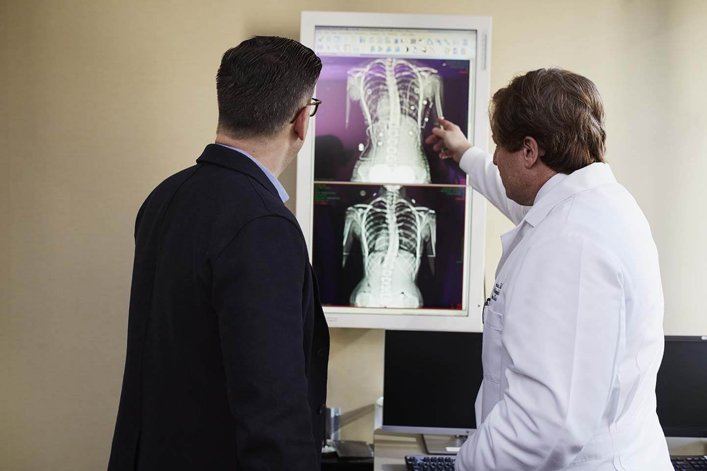
X-ray
X-ray uses a small amount of radiation that passes through the body to quickly capture a single image of your anatomy to assess injury (fractures or dislocations) or disease (bone degeneration). Bones being dense will block the radiation and appear white on the X-ray picture. Specialists will review the pictures and create a report with their findings to aid in diagnosis.
- Assessing injury (see X-ray scan of the hand to the right)
- low-cost, commonly used as the first step